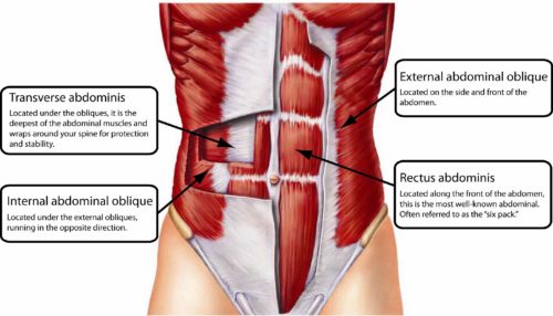
Welcome to the review of day 4 in the cadaver lab. Today’s mission was to work towards the quadriceps and then the abdominal muscles: rectus abdominis, internal and external obliques and transversus abdominus. One of the things you appreciate with this particular dissection is the layering of the abdominals. I wrote about it in the blog post I linked to above but to see it in the body was amazing. It really helped me appreciate the importance of having a strong core but in ALL directions, not just in the up and down (median) plane as most people think (i.e. crunches/rectus abdominus).
We then worked to open the abdomen. This was a bit more challenging because it’s quite different than staying with extremities and dissecting to skeletal muscles. Once we proceeded, it was fascinating to see all the internal organs. The way they all fit together in the abdomen is like a masterful puzzle and you really can appreciate things like how the body adapts to carrying a baby with everything else in there! Being in the lab with 5 other cadavers helped us view the differences between cadavers, which highlighted the differences in body type, shape, size of organs and then we were able to notice differences that led us to surmise about things like health status, conditions the person might have had and things along this line of thought. It’s hard because there was no information about our donors so we did not know conclusively what some of what we saw meant. But it was interesting nonetheless to view the body and make some guesses as to what might have been happening.
The other interesting aspect of dissecting the abdomen was that we could clearly see the psoas muscle. This powerful hip flexor is the one connection between the torso and the lower body and always a bit of a challenge for yoga students. They usually complain it’s “too tight” or “sore” and usually have questions about how to “stretch it.” When you see it, it’s quite a large muscle and even in our donors who were all in advanced stages of life, it was still a very powerful muscle.
The final stage of the day was to view the thoracic cavity, so the heart, lungs, diaphragm and all associated arteries and veins. Let me share that I was, as was everyone else, able to hold a heart in my hand, still attached to the lungs. That was something I will never forget and something that made such an impression on me. To think that surgeons operate on people to address cardiac issues and to even imagine operating on people to do a heart bypass or replacement is amazing once you actually see a “normal” heart and lungs in a cadaver.
One of the other things we were able to do as a side project was further dissect the shoulder of our cadaver so we could look closer at the articulation between the head of the humerus and the glenoid fossa. We had observed before dissection a great deal of crepitis ( joint sounds ) upon movement and we were curious as to the integrity of the connection between the bones. Once we reflected back the pectoralis and the supraspinatus, we were able to better view and manipulate the humerus. We could clearly see the connection was lax and couldn’t quite imagine how our donor managed with that shoulder when she was alive. It also allowed us to view more closely the impact of pushing the head of the humerus forward in weight bearing postures, like Low Push Up, and how indeed it does impinge on all the soft tissue around the joint. This is always a big discussion point among yoga teachers from a safety perspective.
Off to the lab today for my last day of training. My completion certificate is near!

All your days have been filled with some very cool observations. I had no idea the psosa was a big muscle. As you can see I didn’t spell right either. So amazing you are having this experience, and adding to your wealth of knowledge. Enjoy your last day.
Thanks Iris! Yes, the psoas is a big one for sure. It’s a hip muscle but due to its placement, is actually considered part of the core too. Amazing!